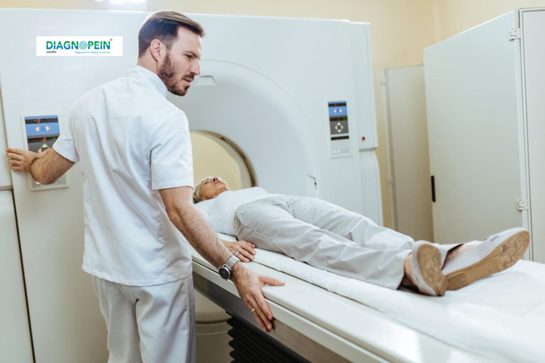
Why MRI Guided Intervention – Nephrostomy with Sialography is Important
MRI-guided nephrostomy with sialography plays a vital role in detecting, localizing, and managing conditions that may not be visible on conventional imaging such as X-ray or ultrasound. MRI provides superior soft tissue resolution, allowing radiologists to guide the catheter with high accuracy, minimize complications, and ensure successful outcomes. Additionally, this imaging method is radiation-free, making it safer for repeated follow-ups.
For patients with obstructed urinary systems due to stones, tumors, or infection, MRI-guided nephrostomy offers targeted drainage of the kidney under direct imaging control. Meanwhile, sialography provides an enhanced view of salivary gland function and ductal structure, helpful in diagnosing chronic infections and ductal stones. Combining these two imaging modalities improves diagnostic confidence and therapeutic planning simultaneously.
Benefits of MRI Guided Nephrostomy with Sialography
-
High diagnostic accuracy for both urinary and salivary duct pathologies
-
Reduced radiation exposure compared to CT-guided interventions
-
Real-time MRI guidance ensures precise catheter placement
-
Quick recovery and minimal patient discomfort
-
Ability to perform diagnosis and treatment in a single procedure
-
Suitable for patients with allergies to iodinated contrast agents
-
Enhanced image clarity for evaluating soft tissues and small lesions
At Diagnopein, these benefits translate into personalized care, faster diagnosis, and reliable interventional outcomes. Our advanced MRI systems and trained interventional radiologists ensure patient safety at every step of the procedure.
Step-by-Step Procedure
Preparation
The patient is advised to undergo basic blood and renal function tests before scheduling the procedure. A mild sedative may be administered to aid comfort. The interventional radiologist reviews the MRI sequences to plan the safest needle path.
Procedure
-
The patient is positioned on the MRI table under sterile conditions.
-
MRI scanning begins to localize the target area within the kidney and salivary gland region.
-
Under continuous MRI visualization, a fine needle is inserted into the affected kidney for nephrostomy or into the salivary duct for sialography.
-
Once the catheter is placed, contrast material suitable for MRI is injected to visualize the ductal system in detail.
-
Drainage or intervention is performed as required under real-time guidance.
Post-Procedure
After the study, images are reviewed by the radiologist for final reporting. The patient is monitored briefly for stability before discharge. Most patients resume normal activity within 24 hours.
MRI Parameters and Imaging Details
MRI-guided nephrostomy and sialography typically use high-resolution T1 and T2 sequences to assess tissue contrast and fluid levels. Dynamic contrast-enhanced imaging helps in delineating ducts and cavities. Key parameters include:
-
TR: 400–900 ms
-
TE: 10–25 ms
-
Slice thickness: 3–5 mm
-
FOV: 30–35 cm
-
Matrix size: 256×256 or higher
These parameters provide optimal resolution for identifying ductal structures and guiding interventional tools accurately.
Clinical Applications
MRI-guided nephrostomy with sialography is ideal for patients with:
-
Obstructive uropathy
-
Hydronephrosis or pyonephrosis
-
Blocked or strictured salivary ducts
-
Chronic salivary gland infections
-
Tumor infiltration affecting ducts
-
Post-surgical complications needing precise drainage
Combining nephrostomy and sialography in MRI guidance maximizes diagnostic efficiency and patient comfort in complex multi-organ cases.
Why Choose Diagnopein
Diagnopein combines innovation and expertise in interventional radiology. Our center is equipped with advanced 3T MRI scanners and sterile interventional suites for image-guided procedures. Every patient receives dedicated attention from a multidisciplinary team focused on achieving accurate diagnosis and safe outcomes. With a commitment to compassionate imaging and excellence, Diagnopein stands as a trusted partner in advanced radiologic interventions.


