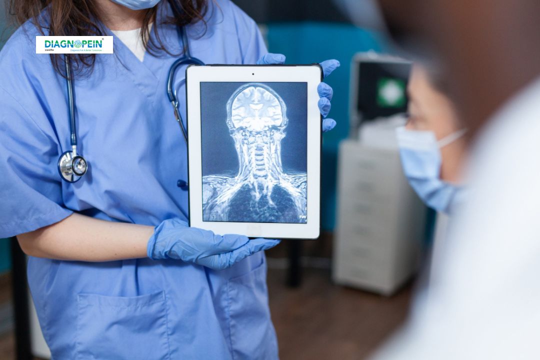
Why MRI Temporomandibular – B/L – With Contrast Is Recommended
The temporomandibular joint connects your jaw to your skull and allows essential movements like talking, chewing, and yawning. When this joint becomes injured or inflamed, it can cause significant discomfort and affect daily life. TMJ MRI scan in Karad helps detect causes of jaw dysfunction accurately.
MRI with contrast is particularly useful when your doctor suspects:
-
Inflammation in the joint structures
-
Disc displacement or internal derangement
-
Degenerative joint disease or arthritis
-
Post-surgical complications
-
Tumors, cysts, or vascular abnormalities around the joint
Contrast dye improves tissue differentiation, enabling our radiologists to see structural and vascular details and detect even minor abnormalities. This ensures that every MRI Temporomandibular joint both sides Karad exam provides maximum diagnostic insight for personalized care.
Patients experiencing jaw stiffness, clicking sounds, or chronic TMJ pain will benefit most from an Advanced MRI for TMJ pain Karad, enabling them to take timely medical action.
Benefits of MRI Temporomandibular – B/L – With Contrast
Choosing an MRI scan for temporomandibular disorder Karad offers multiple benefits, including:
-
High Diagnostic Precision: MRI imaging captures soft tissue details, unlike X-ray or CT scans that mainly visualize bone structures.
-
Comprehensive Assessment: Bilateral scanning ensures both sides of the TMJ are examined simultaneously for comparison.
-
Early Detection: Identifies subtle changes in joint cartilage, ligaments, and surrounding tissues before symptoms worsen.
-
Accurate Treatment Planning: Helps dentists, surgeons, and physiotherapists plan appropriate interventions or therapies.
-
Safe & Non-Invasive: MRI uses no radiation and the contrast material is generally well-tolerated.
At Diagnopein, our commitment to patient safety and accuracy makes us the Best diagnostic center for TMJ MRI in Karad. Every TMJ MRI scan is interpreted by experienced radiologists who ensure precise image evaluation and report generation.
How the MRI TMJ B/L With Contrast Test Is Performed
When you visit Diagnopein for your Contrast MRI for jaw joint Karad, you’ll undergo a quick and comfortable process:
-
Preparation: You may be asked to remove metal objects and avoid eating for a few hours before the scan.
-
Positioning: The radiology technologist positions you on the MRI table; a head coil is used to stabilize your jaw during the scan.
-
Contrast Administration: A small amount of contrast agent is injected into a vein to enhance image detail.
-
Scanning Process: The MRI scanner captures high-resolution images of both TMJs in various mouth positions (open, closed, semi-open).
-
Duration: The entire process usually takes 30–45 minutes.
-
After Scan: You can resume normal activity immediately unless instructed otherwise.
Our MRI parameters are set to a high spatial resolution with thin-slice imaging and contrast enhancement to achieve detailed visualization.
Scan parameters typically include:
-
T1 and T2 weighted sequences
-
Proton density images
-
Oblique sagittal and coronal planes
-
Post-contrast fat-suppressed sequences
These parameters ensure optimal evaluation of joint alignment, disc morphology, and inflammatory changes.
Why Choose Diagnopein Karad for TMJ MRI
Diagnopein provides advanced MRI facilities equipped with modern scanners and expert radiologists specializing in head and neck imaging. We prioritize patient comfort, fast report delivery, and affordable pricing, making us the leading choice for MRI TMJ B/L with contrast Karad and related diagnostic services.
We maintain strict protocols for contrast safety, sterilization, and image accuracy, ensuring every patient receives reliable diagnostic results. Whether you need an MRI Temporomandibular joint both sides Karad for chronic pain or post-surgical evaluation, Diagnopein ensures precision, safety, and care at every step.


