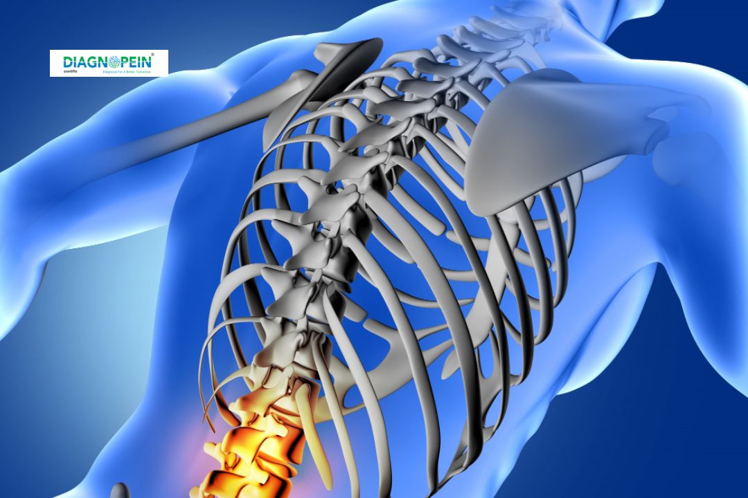
Why MRI Lumbar/ Lumbo-Sacral Spine – With Contrast is Important
An MRI lower back with gadolinium contrast plays a crucial role when conventional MRI images do not provide enough diagnostic clarity. The contrast dye highlights specific spinal structures and abnormalities, enabling the radiologist to distinguish between active inflammation, scar tissue, and new disease lesions.
Key clinical uses include:
-
Evaluating herniated or degenerated discs.
-
Detecting nerve impingement or spinal stenosis.
-
Diagnosing spinal tumors, infections, or cysts.
-
Assessing post-operative changes after back surgery.
By providing clearer insights, an enhanced MRI lumbar spine for herniated disc detection helps doctors plan targeted therapies and avoid unnecessary interventions. It is a safe and highly reliable imaging method recommended by specialists when deeper evaluation of spinal pathology is required.
Benefits of MRI Lumbar/ Lumbo-Sacral Spine with Contrast
Undergoing a Lumbosacral MRI imaging with contrast offers multiple benefits:
-
High Diagnostic Accuracy: The contrast improves visibility of tissues, blood vessels, and inflammation.
-
Early Detection: Detects hidden or active infection and early degenerative changes.
-
Non-invasive and Safe: Uses no harmful radiation. Gadolinium contrast is generally well-tolerated.
-
Better Post-Surgical Evaluation: Distinguishes recurrent disc herniation from scar tissue.
-
Comprehensive View: Visualizes both structural and vascular aspects of the lumbar spine.
At Diagnopein, patients benefit from advanced MRI technology and experienced radiologists who ensure comfort, precision, and safety throughout the scan.
How the MRI Lumbar/ Lumbo-Sacral Spine – With Contrast Test is Done
The scan procedure for MRI Lumbo-Sacral Spine Scan with Contrast near Karad is simple and takes about 30–40 minutes:
-
You will be asked to lie down on the MRI table.
-
A small intravenous line (IV) will be placed for contrast injection.
-
The gadolinium contrast agent is injected midway through the scan.
-
The MRI machine captures detailed cross-sectional images of the lumbar and sacral areas.
-
Radiologists assess the enhanced images for any spinal abnormalities or nerve compression.
You can resume normal activities immediately after the scan. For patients with a history of allergy or kidney issues, a preliminary screening ensures complete safety.
MRI Parameters and Technical Details
Modern MRI systems use advanced imaging protocols for accuracy and safety. Common scan parameters at Diagnopein include:
-
Magnet Strength: 1.5T to 3.0T for high-resolution imaging.
-
Scan Plane: Axial, sagittal, and coronal images.
-
Slice Thickness: 3–4 mm for precise detail.
-
Contrast Used: Gadolinium-based agent for tissue differentiation.
-
Duration: Approximately 30–40 minutes including contrast phase.
The result is a sharp, dynamic image of your lumbar and sacral spine, revealing structural and pathological details essential for diagnosis and follow-up treatment planning.


