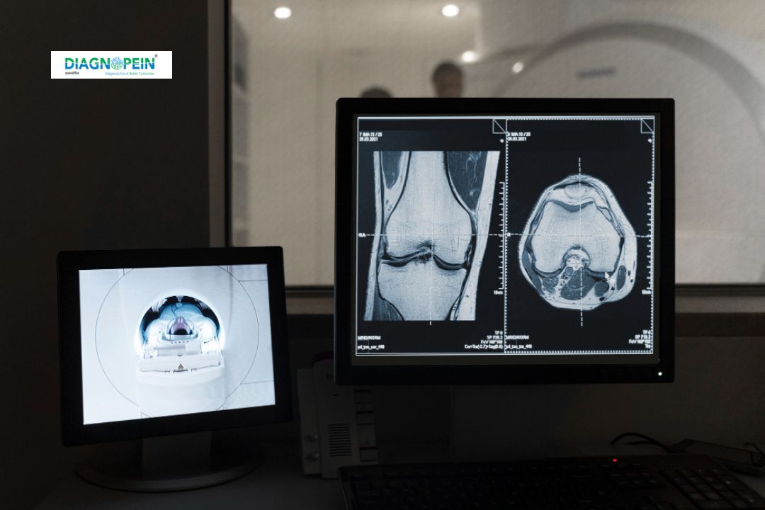
Why MRI Hip with Contrast and Sialography are Important
Combining MRI Hip with contrast and Sialography adds substantial diagnostic value. Contrast material (gadolinium-based) enhances visibility of soft tissue structures that are otherwise indistinct, helping differentiate between healthy and diseased tissue.
Sialography assesses salivary duct and gland function, while MRI Hip detects structural and vascular issues in the hip joint. When done together, they assist in evaluating conditions linked to autoimmune or systemic infections that involve both glandular and joint regions, such as:
-
Rheumatoid arthritis with glandular involvement
-
Chronic joint pain with systemic inflammation
-
Suspected avascular necrosis or synovitis
-
Hip labral tear, cartilage lesion, or tendon injury
-
Post-surgical follow-up and treatment monitoring
This combination also supports surgeons and rheumatologists in planning targeted therapies based on a complete image of musculoskeletal and connective tissues.
Benefits of MRI Hip with Contrast and Sialography
-
High-Resolution Visualization: Offers a detailed view of joint structures, ligaments, and surrounding soft tissues.
-
Contrast Enhancement: Highlights active inflammation, tumor margins, or infection in the hip region.
-
Non-Invasive and Radiation-Free: Unlike CT scans, MRI uses magnetic fields to produce images safely.
-
Functional Assessment: Sialography when integrated with MRI provides a diagnostic edge for evaluating secretion pathways and ductal blockages.
-
Early Diagnosis: Detects conditions before major symptoms develop, improving treatment outcomes.
Patients benefit from quicker diagnosis, better image clarity, and an accurate understanding of both musculoskeletal and glandular pathologies. This multi-dimensional evaluation at Diagnopein aids faster recovery planning and follow-up monitoring.
How the MRI Hip with Contrast and Sialography Test is Done
The test is done under the supervision of certified radiologists and MRI technologists at Diagnopein.
-
Preparation:
Patients should avoid metallic items and inform the team about existing implants or allergies. Fasting for a few hours before contrast administration may be required.
-
Contrast Injection:
A gadolinium-based contrast agent is administered intravenously to highlight vascular and tissue characteristics. For Sialography, a small amount of contrast is introduced into the salivary duct.
-
Imaging Process:
The patient lies on the MRI table, which slides inside the scanner. Multiple sequences are captured in T1, T2, and STIR modes before and after contrast injection. Scanning typically takes 30–45 minutes.
-
Post-Procedure:
Normal activities can be resumed unless otherwise advised. Images are reviewed and reported by senior radiologists, and results are shared digitally.
Key MRI and Sialography Parameters
MRI Hip Parameters
-
Sequence Types: T1, T2, STIR, PD-Fat Sat
-
Slice Thickness: 3–4 mm
-
Field of View (FOV): 22–24 cm
-
Matrix: 256 x 256
-
Contrast Dose: 0.1 mmol/kg body weight (Gadolinium-based)
Sialography Parameters
-
Contrast Type: Iodinated or water-soluble contrast
-
Volume: 0.2–0.5 ml per duct
-
Imaging Mode: Static and dynamic acquisition with MRI synchronization
This protocol ensures maximum visibility of fine glandular details and hip joint anatomy while maintaining patient comfort.
Why Choose Diagnopein
-
Advanced 3T MRI scanners for detailed contrast imaging
-
Expert radiologists specialized in musculoskeletal diagnosis
-
Integration of MRI and Sialography under one roof
-
Digital result delivery and quick turnaround time
-
Patient-focused testing environment with trained technicians
At Diagnopein, every MRI Hip with Contrast and Sialography test is performed with precision, ensuring reliable, accurate, and timely results for clinical decisions.


