Why MRI Head – With Contrast with Sialography is Important
MRI Head – With Contrast with Sialography offers several key advantages in accurate diagnosis. It not only focuses on the brain and cranial structures but also extends to the salivary glands, which can be affected by infections, stones, tumors, or inflammatory conditions.
This dual imaging technique helps clinicians in:
-
Detecting brain lesions, tumors, cysts, or demyelinating diseases
-
Evaluating blood vessels for aneurysms or vascular malformations
-
Identifying inflammation or infection in the salivary ducts
-
Diagnosing autoimmune diseases such as sarcoidosis and Sjögren’s syndrome
-
Planning surgical or medical treatment with high precision
Unlike CT scans, MRI does not use ionizing radiation, making it safe for most patients while offering superior detail in soft tissues.
Benefits of MRI Head – With Contrast with Sialography
The combination of contrast MRI and sialography provides a comprehensive diagnostic advantage. Patients benefit from:
-
Enhanced diagnostic clarity through contrast-enhanced imaging for detecting even minute structural changes.
-
Early disease detection, improving treatment outcomes in conditions like multiple sclerosis, tumors, or vascular disorders.
-
Precise salivary gland visualization, helping detect obstructions or stones in ducts.
-
Non-invasive and safe technique without radiation exposure.
-
Comprehensive head and neck evaluation, saving time and avoiding multiple separate tests.
This advanced modality provides physicians with deep anatomical insights and accurate disease mapping—crucial for effective treatment strategy.
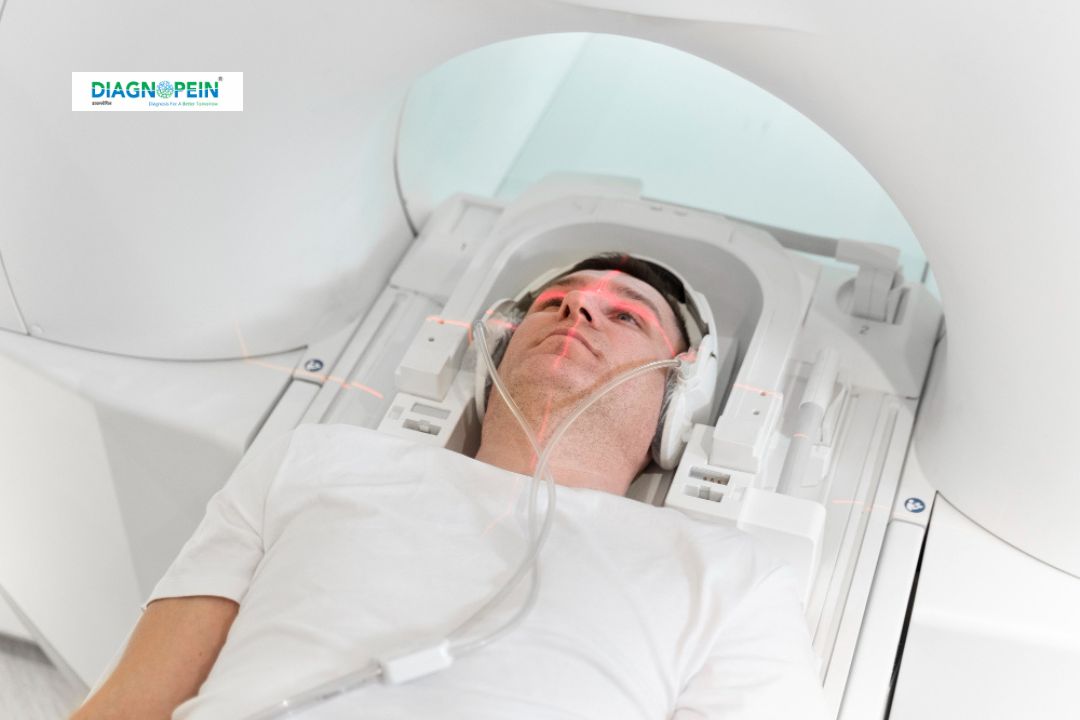
How MRI Head – With Contrast with Sialography is Done
Before the scan, patients are advised to remove metallic objects and fast for a few hours if required. The procedure includes these steps:
-
The patient lies on the MRI table, which slides into the scanner.
-
A contrast dye (gadolinium) is injected intravenously.
-
The MRI machine captures a series of high-resolution images of the brain and head.
-
For the sialography portion, detailed imaging of the salivary glands and ducts is performed using contrast visualization.
-
The entire scan usually takes 45–60 minutes, and the patient can resume normal activities soon after.
The test is painless, though some patients may experience mild warmth or tingling when the contrast agent is injected. Radiologists at Diagnopein analyze these images to prepare an accurate and comprehensive report for your doctor.
MRI Head – With Contrast with Sialography Parameters
Common scan parameters used in MRI Head with contrast with sialography include:
-
Field strength: 1.5 Tesla or 3 Tesla MRI systems
-
Slice thickness: 3–5 mm for high-resolution detail
-
Contrast agent: Gadolinium-based intravenous contrast
-
Sequences: T1-weighted (pre- and post-contrast), T2-weighted, FLAIR, diffusion, and sialographic sequences
-
Scan duration: Approximately 45–60 minutes depending on patient condition
At Diagnopein, our radiology team uses optimized imaging protocols to ensure maximum diagnostic accuracy with patient comfort.