Why MRI Head Scan with Sialography is Important
MRI Head Scan with Sialography plays an essential role in identifying structural, vascular, and glandular problems that routine imaging might miss. It is particularly valuable for detecting:
-
Salivary gland blockages or inflammation (sialadenitis)
-
Tumors or cysts in salivary glands or ducts
-
Neurological conditions such as brain tumors, aneurysms, or demyelinating diseases
-
Trauma or post-surgical complications involving facial or head regions
The integration of MRI technology with advanced Sialography enhances clarity and precision, allowing physicians to differentiate between soft tissue pathologies, glandular lesions, and neural abnormalities. This technique is often preferred over CT scans because it avoids radiation exposure and delivers superior soft tissue contrast.
Benefits of MRI Head Scan with Sialography
The test offers several key advantages for accurate diagnosis and patient safety:
-
High-resolution imaging: Produces clear and detailed views of soft tissues, glands, and brain structures.
-
Non-invasive and radiation-free: Uses powerful magnetic fields and radio waves instead of harmful ionizing radiation.
-
Comprehensive diagnosis: Evaluates both neurological and salivary systems in a single session.
-
Early detection: Identifies diseases in their earliest stages for better clinical outcomes.
-
Guided treatment planning: Helps surgeons and specialists plan precise interventions, especially in complex cases involving head or facial regions.
Because of its proven safety and diagnostic accuracy, MRI Head Scan with Sialography is one of the most trusted imaging options for ENT, dental, and neurological assessments.
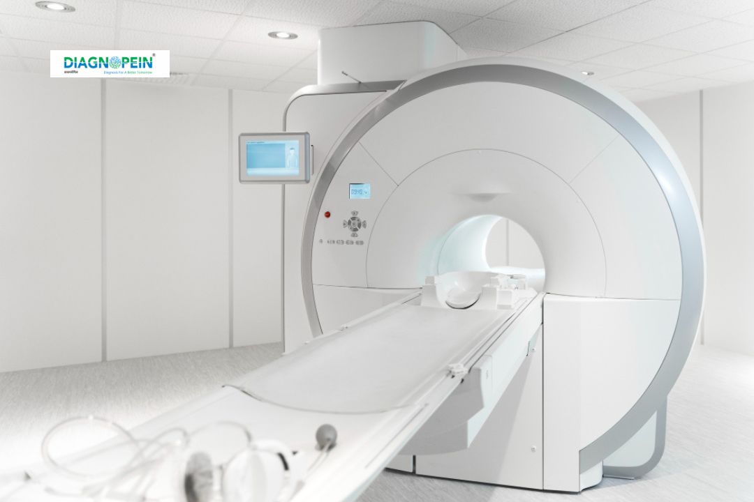
How the MRI Head Scan with Sialography is Performed
-
Patient Preparation:
The patient is asked to remove all metal objects and lie down on the MRI table. In some cases, a contrast agent may be used to enhance visualization of the salivary ducts.
-
MRI Procedure:
The scanner creates detailed images using magnetic fields. The patient must remain still during the process, which usually takes 30 to 45 minutes.
-
Sialography Process:
A mild contrast solution is introduced into the salivary duct to map its structure. This dye is safe and quickly excreted from the body.
-
Image Analysis:
Radiologists analyze both MRI and Sialography images to identify any structural changes or blockages, ensuring a comprehensive evaluation.
Patients experience minimal discomfort, and results are typically available within 24 to 48 hours. The procedure is safe for most individuals, including those requiring repeated imaging.
Typical Scan Parameters
-
Field strength: 1.5 to 3 Tesla MRI scanner
-
Sequence types: T1-weighted, T2-weighted, STIR, and diffusion imaging
-
Slice thickness: 3–5 mm for high-resolution detailing
-
Contrast agent: Gadolinium-based contrast for enhanced salivary visualization (when necessary)
-
Duration: Approximately 40–60 minutes depending on complexity
These parameters ensure detailed imaging of both neural and glandular structures for maximum diagnostic accuracy.
When to Consult Your Doctor
Consult a healthcare professional if you experience persistent facial pain, recurrent swelling in the jaw region, difficulty swallowing, or any neurological symptoms. An MRI Head Scan with Sialography helps pinpoint the root cause of such conditions quickly and effectively.