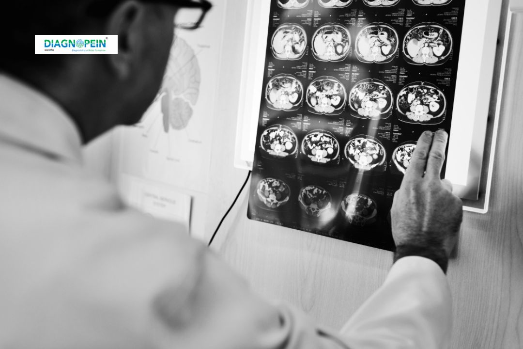
What is MRI Head/Brain – Without Contrast with Sialography?
MRI (Magnetic Resonance Imaging) Head or Brain – Without Contrast uses strong magnetic fields and radio waves to produce clear and detailed images of the brain’s soft tissues and nervous system. Sialography is an imaging technique focused on the salivary glands and their ducts, helping detect blockages, infections, or inflammation.
When both scans are performed together, they provide a comprehensive evaluation of the brain and facial regions—crucial in identifying neurological and glandular conditions in a single session. This non-invasive test avoids radiation and offers superior clarity compared to CT or X-ray imaging.
Why is MRI Head/Brain – Without Contrast with Sialography Important?
MRI Head/Brain without contrast combined with Sialography helps detect, evaluate, and monitor a wide range of conditions affecting both the central nervous system and salivary structures.
Key diagnostic uses include:
-
Early detection of brain lesions, tumors, or cysts.
-
Evaluation of headaches, dizziness, seizures, and neurological symptoms.
-
Diagnosis of salivary gland disorders like sialolithiasis (stones), infections, or ductal obstructions.
-
Assessment of facial nerve pathologies and post-surgical or trauma cases.
The technique is particularly important for patients where contrast-based MRI is contraindicated, ensuring safety and diagnostic accuracy.
Benefits of MRI Head/Brain – Without Contrast with Sialography
1. Non-invasive and radiation-free:
The scan uses no ionizing radiation, making it safe for adults, children, and patients requiring repeated follow-up imaging.
2. Enhanced image quality:
High-resolution images help radiologists visualize even small structural abnormalities in the brain and salivary ducts.
3. Safe for contrast-sensitive patients:
Ideal for individuals allergic to gadolinium-based contrast agents or those with compromised kidney function.
4. Dual diagnostic coverage:
Combines brain and salivary gland imaging, saving time and cost while offering a complete clinical picture.
5. Early and accurate detection:
Facilitates prompt treatment planning and better outcomes in neurological and glandular disorders.
How the MRI Head/Brain – Without Contrast with Sialography Test is Performed
1. Preparation:
-
No fasting or major preparation needed before the scan.
-
Patients must remove metal objects like jewelry, watches, or hearing aids.
-
Inform the radiologist about any implants or pacemakers.
2. Procedure:
-
The patient lies still on the MRI table, which slides into the scanner tunnel.
-
Multiple series of images are captured using magnetic gradients.
-
Sialography sequences focus on the salivary ducts without radiation or dye injection.
-
The entire process usually takes 30–45 minutes.
3. Post-scan:
Patients can resume normal activities immediately. The report is analyzed by an experienced radiologist and shared with the referring doctor.
Scan Parameters and Technical Specifications
Common MRI Parameters:
-
Repetition Time (TR): 4000–6000 ms
-
Echo Time (TE): 90–120 ms
-
Slice Thickness: 3–5 mm
-
FOV (Field of View): 220–250 mm
Sialography Parameters:
-
TR: 2500–3500 ms
-
TE: 80–100 ms
-
Slice Thickness: 2–3 mm for ductal clarity
These optimized parameters ensure diagnostic-quality images with exceptional detail for both brain and salivary duct evaluation.
Why Choose Diagnopein for MRI Head/Brain – Without Contrast with Sialography?
Diagnopein uses state-of-the-art MRI scanners and advanced Sialography techniques conducted by skilled radiologists and technologists. Our team ensures a comfortable, safe, and precise imaging experience while maintaining quick reporting and professional care.
We prioritize patient safety, clarity of diagnosis, and technological excellence for comprehensive imaging assessments.