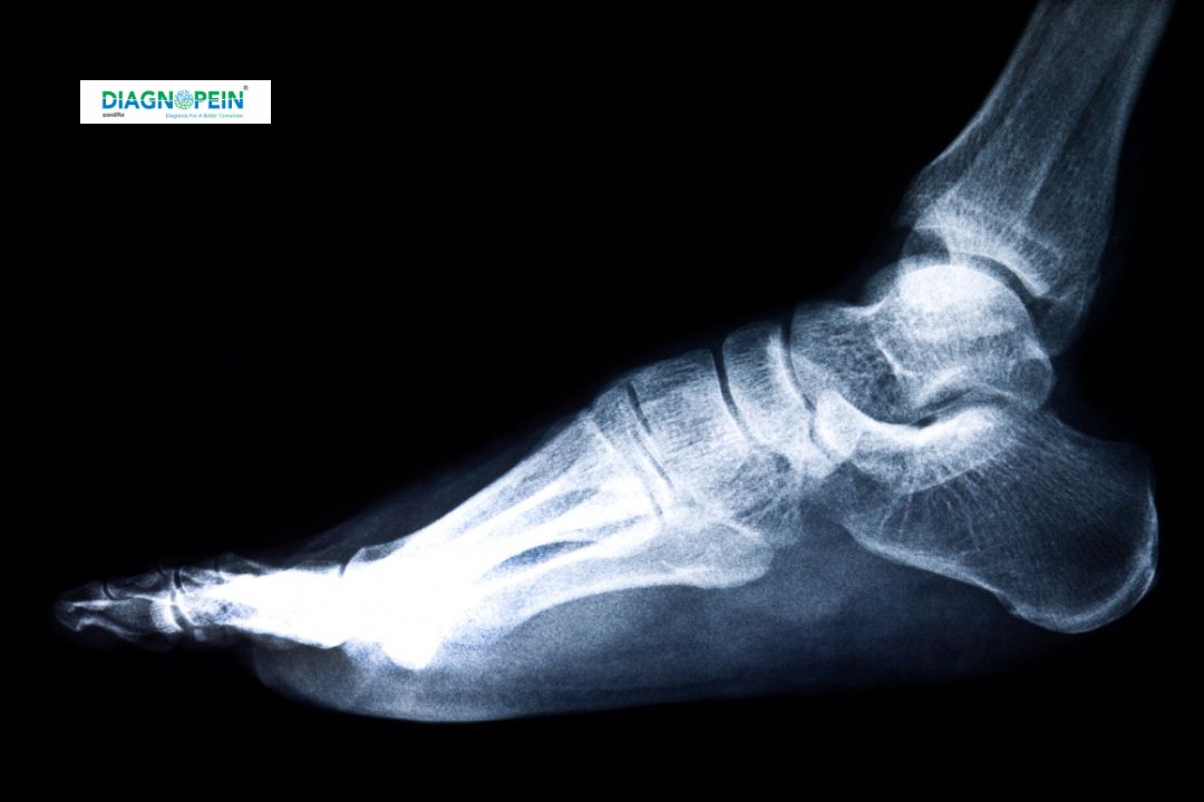
Why MRI Foot is Important
The foot is one of the most complex structures in the human body, made up of 26 bones and numerous soft-tissue connections. Injuries or pain in the foot can significantly affect walking, balance, and mobility. MRI Foot imaging is essential when symptoms such as pain, swelling, or stiffness persist despite basic diagnostic tests.
MRI Foot is commonly recommended for:
-
Chronic heel or ankle pain
-
Sports injuries or ligament tears
-
Suspected fractures not visible in X-rays
-
Infections or abscesses in foot tissues
-
Arthritis or degenerative joint diseases
-
Diabetic foot ulcers and complications
-
Soft tissue masses or suspected tumors
By identifying the root cause accurately, the MRI Foot scan helps doctors prevent further damage and plan effective treatment.
Benefits of MRI Foot Scan
MRI Foot provides several diagnostic advantages that make it one of the most reliable imaging tests for foot and ankle conditions.
-
High-Resolution Imaging: MRI produces high-definition images of soft tissues, joints, and bones without radiation exposure.
-
Early Detection: Detects small fractures, ligament strains, or tendon injuries that might go unnoticed on X-rays or CT.
-
Non-Invasive and Safe: MRI uses strong magnetic fields instead of ionizing radiation, making it safe for repeated use.
-
Guides Surgery and Treatment: Helps surgeons and orthopedic specialists plan precise interventions.
-
Quick and Painless: The procedure typically takes 20–30 minutes and does not require any incisions or recovery time.
At Diagnopein, every MRI Foot scan is performed under the supervision of trained radiology experts to ensure clarity, speed, and comfort.
How MRI Foot Test is Done
The MRI Foot scan is a simple and comfortable procedure conducted in an advanced MRI room. Here’s what happens during the test:
-
Preparation: You will be asked to remove all metal objects, jewelry, or electronic items before the scan. You might be asked to wear a hospital gown.
-
Positioning: You will lie down on the MRI table, with your foot positioned inside the scanner coil. Ear protection is provided to minimize noise from the machine.
-
Scanning: The MRI machine takes multiple high-resolution images while you remain still. You may hear knocking or humming sounds during the process.
-
Contrast (If Required): In some cases, a contrast dye is injected to highlight blood vessels or soft tissue structures for better clarity (e.g., MRI Foot with contrast).
-
Completion: The scan usually takes around 25–30 minutes. Once done, you can resume your normal activities immediately unless sedation is used.
MRI Foot Scan Parameters
-
Magnet Strength: 1.5T or 3T MRI (depending on the case)
-
Slice Thickness: 3–4 mm for detailed visualization
-
Imaging Planes: Axial, sagittal, and coronal
-
Coil Used: Dedicated foot and ankle coil for high image quality
-
Sequence Types: T1, T2, STIR, PD Fat Sat sequences for bone, ligament, and soft tissue evaluation
Each MRI Foot scan is customized based on the patient’s symptoms and physician’s diagnostic requirements.
Why Choose Diagnopein for MRI Foot
Diagnopein is equipped with state-of-the-art MRI technology, ensuring quick, accurate, and comfortable scanning experiences. Our radiologists have deep expertise in musculoskeletal imaging and deliver timely, reliable reports. With affordable pricing, clean facilities, and digital result access, we make diagnostic care more accessible to every patient.