Why MRI Fistulagram is Important
MRI Fistulagram plays a key role in the diagnosis and management of complex fistulas. It provides high-resolution images that allow doctors to:
-
Locate the exact course and opening of the fistula tract
-
Identify any associated abscess or inflammation
-
Evaluate multiple tracts and branching patterns
-
Differentiate between active and healed disease regions
Early detection through MRI helps prevent recurrent infections, unnecessary surgery, and delays in treatment. Surgeons rely on this imaging before performing corrective operations, ensuring better outcomes and minimal complications. In chronic conditions like Crohn’s disease, regular MRI Fistulagrams assist in monitoring disease progression and treatment response.
Benefits of MRI Fistulagram
This imaging test provides several advantages over conventional methods:
-
Non-invasive and painless: No surgical instruments or incisions are required.
-
No radiation exposure: Safe for all age groups, including younger patients needing repeat follow-ups.
-
High diagnostic accuracy: Detects even small or hidden fistula tracts.
-
Supports treatment planning: Radiologists can guide physicians on the best surgical or medical approach.
-
Quick recovery: Since it’s an outpatient scan, patients can resume normal activities immediately after.
MRI Fistulagram is ideal for detecting deep-seated or recurrent fistulas, especially in sensitive pelvic and anorectal regions, where traditional imaging may be limited.
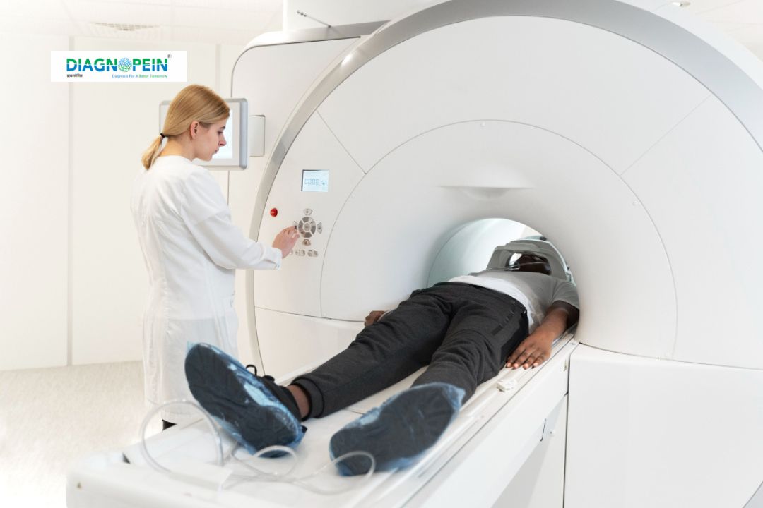
How the MRI Fistulagram Test is Done
The procedure is simple, comfortable, and usually completed within 30–45 minutes.
A radiology technician guides the patient through each step carefully:
-
Preparation: Patients are advised to wear comfortable clothes and remove any metal items. Sometimes a mild enema or fasting may be recommended.
-
Positioning: The patient lies on the MRI table, and depending on the fistula location, a surface coil is placed around the area.
-
Contrast administration (if needed): A small amount of contrast agent may be injected into the tract or through a vein to highlight the fistula clearly.
-
Scanning: The MRI machine generates images as the patient remains still. Technicians may ask the patient to hold their breath briefly during specific sequences.
-
Image interpretation: The radiologist reviews the data and prepares a detailed diagnostic report for the treating doctor.
Scan Parameters and Technical Details
MRI Fistulagram is performed using high-resolution sequences, typically on 1.5 Tesla or 3 Tesla MRI scanners. Standard imaging parameters include:
-
T1-weighted images: Outline anatomical structures
-
T2-weighted images: Highlight inflammation and fluid
-
Fat-suppressed sequences: Differentiate fat from abnormal tissues
-
Post-contrast sequences: Identify active tracts and abscesses
Adjusted slice thickness (2–3 mm) and multi-planar reconstruction ensure complete visualization of fistula tracts and surrounding tissues.
When to Consider an MRI Fistulagram
Your doctor may recommend an MRI Fistulagram if you have:
-
Chronic anal or rectal discharge
-
Previous surgery for fistula but persistent symptoms
-
Crohn’s disease or inflammatory bowel disease
-
Suspected recurrent or complex fistulas
-
Non-healing wounds or infections
Book Your MRI Fistulagram Appointment
At Diagnopein, we use advanced high-field MRI scanners and expert radiology interpretation to provide accurate and detailed MRI Fistulagram reports. Our team ensures patient comfort, precision imaging, and fast reporting to aid effective treatment decisions.
Book your MRI Fistulagram test today for precise diagnosis and expert care.