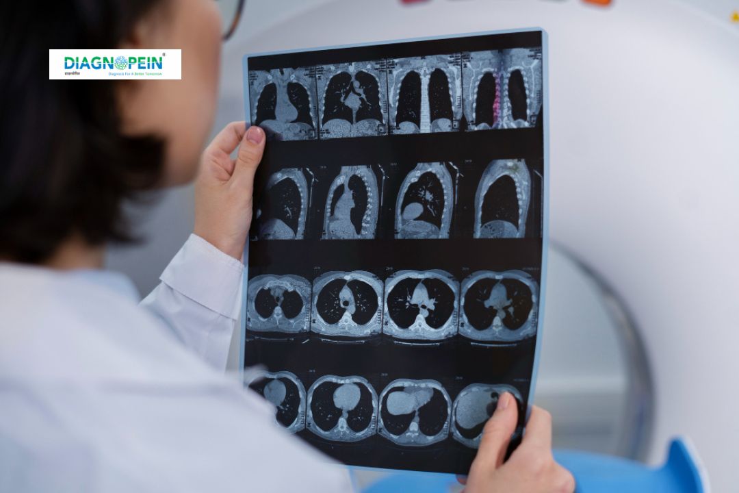
Importance of MRI Extremities – Without Contrast
MRI Extremities – Without Contrast is important for evaluating a wide range of orthopedic and musculoskeletal conditions. It provides detailed visualization of both bone and soft tissue structures, helping clinicians identify causes of pain, restricted movement, or swelling without exposing patients to contrast dye.
It is particularly beneficial for:
-
Detecting ligament or tendon tears, such as ACL or rotator cuff injuries
-
Assessing joint effusion, cartilage damage, and bone marrow edema
-
Identifying early signs of arthritis or osteonecrosis
-
Evaluating tumors, infections, or cystic lesions in the extremities
-
Monitoring postoperative healing and recovery
Because the scan does not require contrast, it eliminates any risk of allergic reaction or kidney-related side effects, making it suitable for a broad range of patients, including children and those with renal impairment.
Benefits of MRI Extremities – Without Contrast
MRI extremity scans offer multiple advantages to patients and clinicians:
-
Detailed Soft Tissue Imaging: Provides superior visualization of ligaments, muscles, and cartilage compared to other imaging methods.
-
Safe and Painless: No radiation exposure or need for injections.
-
Early Detection: Identifies conditions before they progress, aiding in effective treatment planning.
-
Non-Invasive Diagnostic Option: Helps avoid exploratory surgeries by offering clear internal imaging.
-
Shorter Scan Time: Advanced MRI technologies allow quicker examinations and improved patient comfort.
This imaging method is often preferred for follow-up scans, ongoing injury assessments, and evaluations before or after orthopedic surgeries.
How the Test is Done
The MRI extremity scan is simple, safe, and typically completed within 30 to 45 minutes. The process includes:
-
Preparation: You may be asked to remove metallic objects and change into a hospital gown. No special fasting or medication adjustments are required.
-
Positioning: The patient lies comfortably on the MRI table. Only the extremity being examined (arm or leg) is placed inside the MRI coil.
-
Imaging: The machine uses magnetic and radio waves to create image slices that are processed into 3D visuals. You must remain still to get clear results.
-
Completion: Once the scan is done, images are reviewed by a radiologist, and diagnostic reports are provided to your doctor for further management.
The test is completely painless and involves no injections or radiation.
Technical Parameters
Typical parameters for MRI Extremities – Without Contrast may include:
-
Field strength: 1.5T or 3.0T
-
Sequence types: T1, T2, STIR, and Proton Density (PD)
-
Slice thickness: 3–5 mm depending on the region
-
Coil types: Dedicated extremity or surface coils for enhanced resolution
-
Scan duration: 30–45 minutes on average
These optimized parameters ensure excellent image clarity and diagnostic value for clinicians.


