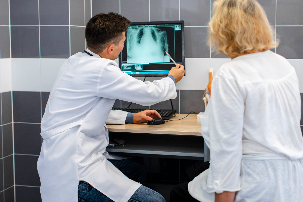
MRI Chest – Without Contrast
Magnetic Resonance Imaging (MRI) of the chest without contrast is a powerful diagnostic technique that provides high-resolution images of the chest region, including the lungs, heart, mediastinum, blood vessels, and chest wall structures. This non-invasive scan helps physicians detect, monitor, and evaluate a wide range of thoracic diseases without using contrast dye, making it safer for patients with contrast allergies or kidney conditions.
Why MRI Chest – Without Contrast is Important
MRI Chest Without Contrast is essential when there is a need for high-quality imaging in patients with sensitivity to gadolinium-based contrasts or with impaired renal function. It provides significant diagnostic value in certain thoracic conditions while avoiding the potential risks of allergic reaction or nephrogenic systemic fibrosis associated with contrast-enhanced scans.
This scan helps to:
-
Differentiate between benign and malignant chest lesions.
-
Monitor progression or regression of chest wall or lung diseases.
-
Evaluate mediastinal masses and heart abnormalities.
-
Offer cross-sectional imaging insights without radiation exposure.
-
Serve as a follow-up modality for patients post-surgery or therapy.
By focusing exclusively on signal differences between tissues, it provides reliable anatomical information that can guide further diagnostic or therapeutic decisions.
Benefits of MRI Chest – Without Contrast
MRI Chest Without Contrast offers numerous benefits for both patients and clinicians:
-
Safe and non-invasive: No radiation or contrast injection involved.
-
Kidney-safe imaging: Suited for patients with renal impairment.
-
Excellent soft tissue clarity: Distinguishes between soft tissues better than CT scans in many cases.
-
Allergy-free option: Ideal for patients allergic to iodine or gadolinium contrast.
-
Superior multiplanar imaging: Provides coronal, sagittal, and axial sections for a comprehensive view.
Physicians often prefer non-contrast MRI for preliminary assessments, long-term disease monitoring, or follow-up after surgery when the area of interest is already known.
How the MRI Chest – Without Contrast Test is Done
The procedure is simple, comfortable, and typically takes 30–45 minutes. The patient lies flat on an MRI table that slides into the machine. Coils placed around the chest area capture detailed magnetic signals, and a series of sequences are performed to visualize the organs and tissues.
Before the scan:
-
Remove all metallic objects or accessories.
-
Change into a hospital gown.
-
Patients must remain still for clear imaging.
-
Breathing instructions may be given during the scan for better diaphragm and lung capture.
Since no contrast dye is used, there are no injections or associated side effects. After the test, normal activities can be resumed immediately.
MRI Chest – Without Contrast Parameters
Common scan parameters used in this MRI include:
-
Field Strength: 1.5T or 3T MRI scanner
-
Slice Thickness: 3–5 mm for detailed visualization
-
Sequences: T1-weighted, T2-weighted, STIR, and diffusion-weighted sequences
-
Imaging Planes: Axial, coronal, and sagittal
-
Scan Time: Approximately 30–40 minutes
Radiologists tailor these parameters to optimize image quality and diagnostic precision depending on the patient’s condition and clinical focus.