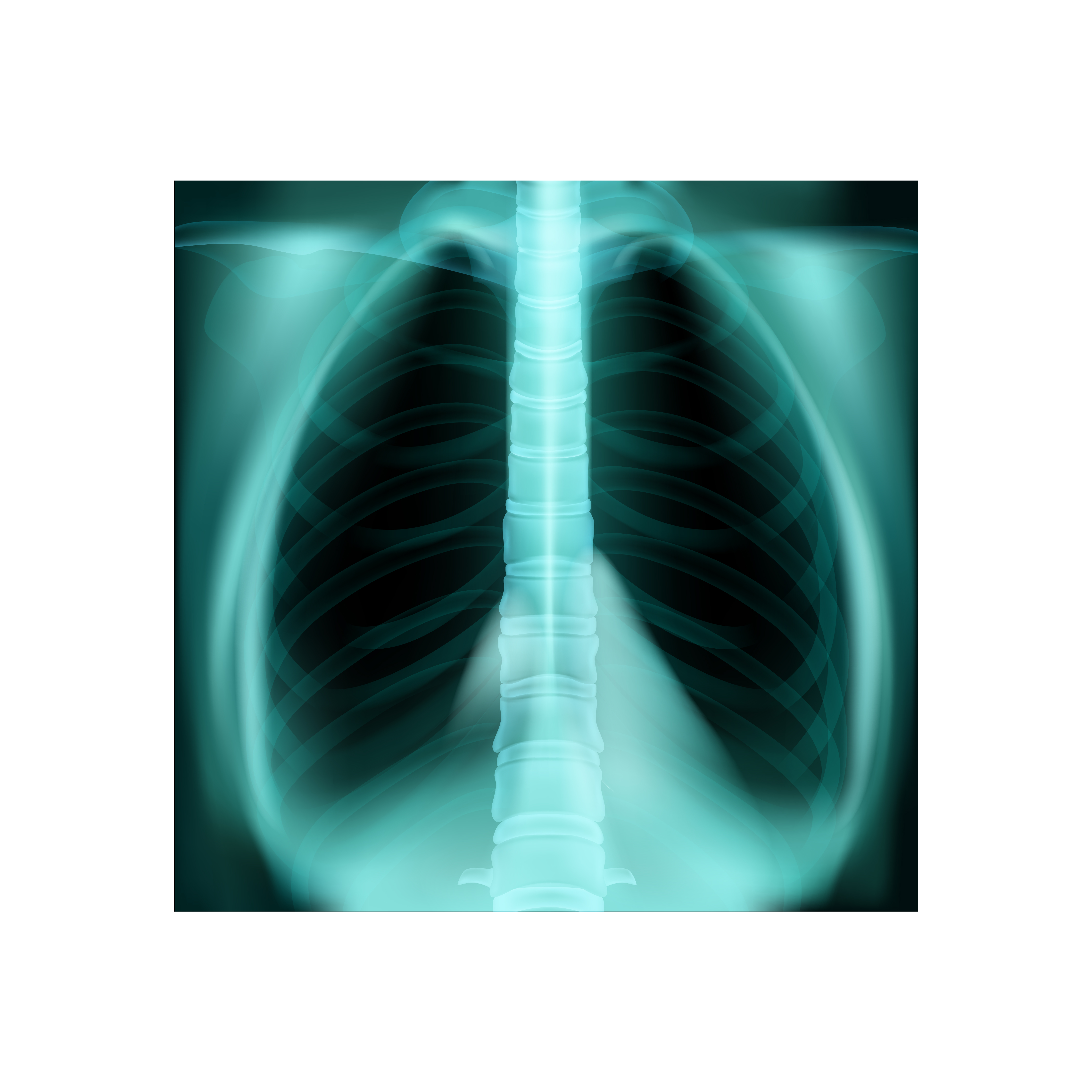
Why MRI Chest – With Contrast is Important
MRI Chest with contrast plays a vital role in diagnosing complex thoracic conditions where other imaging methods, such as CT scans or X-rays, may be limited. The contrast dye increases visualization of soft tissues and blood flow, making it an important tool for accurate assessment.
It is particularly useful to:
-
Detect tumors, cysts, or masses within the chest
-
Evaluate heart and vascular diseases
-
Assess congenital thoracic abnormalities
-
Identify inflammation, infection, or fibrosis
-
Monitor response to ongoing treatment such as chemotherapy
This test is often recommended when doctors need a deeper look beyond basic imaging. For cancer evaluation or cardiovascular assessment, MRI with contrast delivers critical diagnostic insights that can impact treatment outcomes.
Key Benefits of MRI Chest – With Contrast
MRI Chest with contrast offers several advantages over traditional imaging modalities. The detailed and enhanced imaging improves diagnostic precision and patient care.
Main benefits include:
-
High-definition images for better detection of thoracic abnormalities
-
No exposure to ionizing radiation
-
Enhanced tissue distinction between healthy and diseased areas
-
3D imaging capability for accurate anatomical mapping
-
Improved evaluation of vascular and cardiac structures
Modern MRI scanners at Diagnopein ensure patient comfort and faster scan times. The procedure is safe when performed under professional supervision, and allergic reactions to contrast are rare but monitored carefully.
How MRI Chest – With Contrast is Performed
The test usually takes about 30–45 minutes. Patients are advised to avoid food for a few hours before the scan if instructed. During the procedure, a small amount of contrast dye is injected through a vein, followed by the imaging process inside the MRI machine.
Procedure steps:
-
The patient lies down on a sliding MRI table.
-
A contrast agent is administered intravenously.
-
The scanner captures multiple cross-sectional images of the chest.
-
Technologists ensure minimal movement to maintain image quality.
-
After completion, the radiologist analyzes the images and prepares a detailed report.
Patients can usually resume normal activities after the test unless otherwise advised by the physician. Diagnopein’s staff ensures that the process is comfortable, safe, and handled with expert precision.


