Why MRI Contrast is Important
MRI with contrast plays a crucial role in improving diagnostic accuracy. The contrast material travels through the bloodstream, allowing clear visualization of blood flow and tissue structure. This makes it easier to detect problems such as cancer, multiple sclerosis, or vascular diseases at their earliest stages.
Importance of MRI contrast includes:
-
Detects lesions and small tumors not visible on standard MRI scans.
-
Differentiates between active and inactive disease regions.
-
Helps assess organ function, tissue perfusion, and inflammation levels.
-
Aids surgeons in preoperative planning and monitoring post-treatment progress.
Without the use of contrast, many internal abnormalities can remain hidden. At Diagnopain, our advanced MRI systems ensure optimal contrast distribution for precise image capture and reliable diagnosis.
Benefits of MRI Contrast Scans
MRI contrast scans offer numerous clinical benefits over routine MRI procedures. The imaging quality and diagnostic yield are significantly improved due to the highlighting of pathological tissues.
Key benefits include:
-
Enhanced Image Clarity: Clearer visualization of blood vessels and soft tissues.
-
Early Disease Diagnosis: Detects abnormalities at an earlier stage, improving treatment outcomes.
-
Non-invasive Evaluation: No radiation exposure; uses magnetic fields and radio waves only.
-
Comprehensive Assessment: Effective for evaluating tumors, infections, and vascular blockages.
-
Monitoring Treatment Response: Useful in tracking healing or disease progression after therapy.
Patients undergoing MRI contrast scans experience quick results with high accuracy, enabling clinicians to make confident diagnostic decisions.
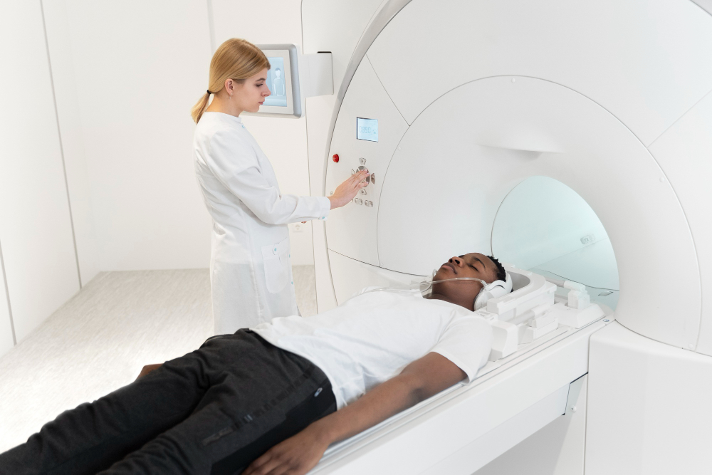
How MRI Contrast Testing is Performed
At Diagnopain, the MRI contrast process is handled with utmost safety and precision. Patients are first screened for allergies, kidney function, and medical history to ensure suitability for contrast use.
Procedure Steps:
-
The patient is positioned comfortably on the MRI table.
-
A gadolinium-based contrast agent is administered through an intravenous (IV) line.
-
The MRI scanner uses magnetic waves to capture detailed images of the internal organs.
-
Patients are monitored throughout for any rare side effects or discomfort.
-
The radiologist analyzes the enhanced images for diagnostic interpretation.
The entire scan usually takes between 30–60 minutes, depending on the body part being examined. After the test, patients can resume their normal activities unless otherwise advised by the radiologist.
MRI Contrast Parameters
At Diagnopain, MRI contrast imaging is tailored using advanced parameters that ensure the best image quality for different organs:
-
Field Strength: 1.5T or 3T MRI units depending on diagnostic need.
-
Contrast Agent Type: Gadolinium-based (macrocyclic and linear agents available).
-
Dosage: Adjusted to patient weight and type of study.
-
Sequences Used: T1-weighted, T2-weighted, FLAIR, and diffusion-weighted imaging.
-
Post-Processing: 3D reconstruction and contrast kinetics analysis for enhanced visualization.
Accurate parameter selection minimizes artifacts and enhances tissue distinction, ensuring reliable diagnostic outcomes across all MRI studies.