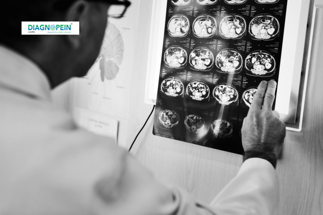
Why MRI Brain Parkinson Protocol is Needed
Diagnosing Parkinson’s disease clinically can be challenging in early stages. MRI Brain Parkinson protocol helps clinicians detect early neurodegenerative changes even before classical symptoms fully appear. It acts as a valuable complement to neurological assessments by offering visual evidence of underlying pathology.
Common reasons for prescribing this scan include:
-
Differentiating idiopathic Parkinson’s disease from Parkinson-plus syndromes.
-
Ruling out secondary causes of parkinsonism such as vascular lesions or structural abnormalities.
-
Monitoring disease progression and response to therapy.
-
Investigating unexplained tremors or rigidity.
This non-invasive imaging tool assists doctors in confirming clinical suspicion with high accuracy and in planning precise treatment strategies based on MRI findings.
Benefits of MRI Brain Parkinson Protocol
MRI Brain Parkinson scanning provides multiple diagnostic and research advantages. The resulting images give valuable insight into both anatomy and microstructural integrity.
Key benefits include:
-
Early and accurate detection of Parkinson’s disease.
-
Ability to visualize changes in substantia nigra and basal ganglia.
-
Non-invasive evaluation with no radiation exposure.
-
Helps differentiate Parkinson’s from similar neurodegenerative disorders.
-
Supports neurologists in tracking disease progression and treatment response.
-
Enhances clinical confidence in diagnosis through objective imaging evidence.
Diagnopein’s cutting-edge MRI systems, along with tailored sequences for Parkinson evaluation, ensure reliability, precision, and consistency in diagnostic results.
MRI Testing Procedure and Parameters
At Diagnopein, the MRI Brain Parkinson protocol is performed under the supervision of experienced radiologists and technologists. The procedure is safe, quick, and comfortable for patients.
Procedure steps:
-
The patient is positioned comfortably in the MRI scanner.
-
High-field MRI (preferably 3 Tesla) is used for best resolution.
-
Specific Parkinson sequences are acquired, including:
-
T1-weighted and T2-weighted imaging
-
Susceptibility-weighted imaging (SWI)
-
Diffusion Tensor Imaging (DTI)
-
Neuromelanin-sensitive sequences
-
Iron quantification techniques (R2*, QSM)
-
The whole scanning session usually takes 30–45 minutes.
-
Images are processed and analyzed for signs of nigral signal loss, asymmetry, or abnormal iron accumulation.
Common parameters:
-
Field strength: 3.0 Tesla preferred
-
Voxel size: 1 mm or lesser for fine detail
-
Slice thickness: 1–3 mm for clear structure definition
-
Orientation: axial and coronal planes through midbrain
-
DTI b-values: 0 and 1000 s/mm² for white matter tract analysis
After the scan, detailed images are reviewed by neuro-radiologists for quantitative and qualitative findings. Reports highlight changes in substantia nigra integrity, midbrain patterns, and diffusion metrics used to support Parkinson’s diagnosis.


