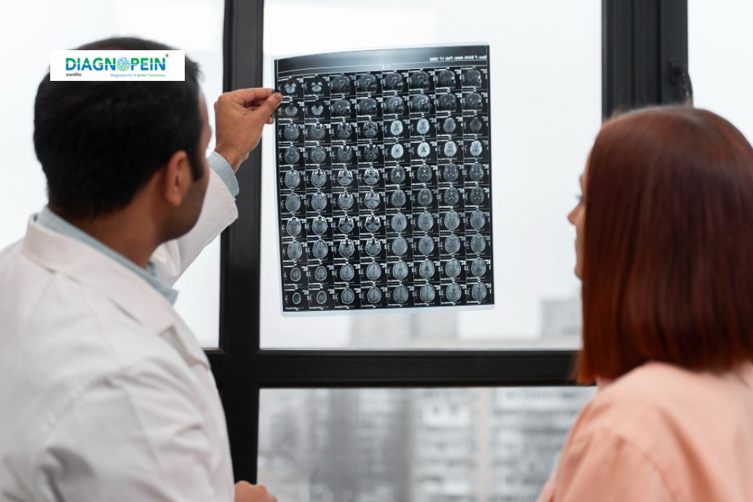
Best MRI Brain with Contrast
Magnetic Resonance Imaging (MRI) of the brain with contrast is an advanced diagnostic test that provides detailed pictures of the brain’s structure, blood vessels, and tissues. By using a contrast dye (commonly gadolinium-based), radiologists can differentiate normal and abnormal areas in the brain more accurately. At Diagnopein, we use high-resolution MRI technology and expert radiologists to ensure early and precise detection of neurological conditions.
Overview of MRI Brain with Contrast
MRI Brain with contrast involves injecting a special contrast agent into a vein before scanning. This dye enhances the visibility of brain tissues and blood flow on the images. It helps detect abnormalities not visible in non-contrast MRIs. The procedure is safe, non-invasive, and provides comprehensive brain visualization without radiation exposure.
Common conditions evaluated include:
-
Brain tumors and metastases
-
Multiple sclerosis (MS) lesions
-
Infections or brain abscesses
-
Stroke and brain ischemia
-
Inflammatory or demyelinating diseases
-
Blood-brain barrier disruptions
-
Post-surgical evaluation of the brain
At Diagnopein, our MRI systems produce high-resolution images using optimized contrast sequencing, providing a reliable view of even minute abnormalities.
Why MRI Brain with Contrast is Recommended
A standard MRI scan detects structural issues, but adding contrast improves diagnostic accuracy by highlighting tissue differences and vascular leaks. This technique is particularly helpful when physicians suspect:
-
Brain tumors, cysts, or metastasis
-
Inflammation or infection of the meninges
-
Blood vessel malformations or aneurysms
-
Post-surgical brain evaluation to differentiate scar tissue from active disease
-
Conditions like multiple sclerosis where contrast enhancement shows active lesion sites
MRI Brain with contrast is recommended when precise tissue characterization is necessary for treatment planning or monitoring therapy response.
Benefits of MRI Brain with Contrast
MRI Brain with contrast provides several diagnostic and clinical advantages:
-
Enhanced detection: Highlights tumors, lesions, or abnormal brain tissues not visible on plain MRI.
-
Accurate differentiation: Helps separate active disease areas from old or inactive ones, especially in multiple sclerosis or post-surgery cases.
-
Better vascular imaging: Shows blood vessel abnormalities, hemorrhages, or blockages more distinctly.
-
Precise treatment planning: Allows doctors to determine exact lesion locations for surgery or radiation therapy.
-
Superior monitoring: Tracks response to therapy and progression of neurological diseases.
By choosing Diagnopein, patients benefit from expert supervision, faster scan times, and advanced 3T MRI systems designed for detailed brain mapping.
Testing Procedure and Scan Parameters
Procedure Steps
-
Preparation: You may be asked to avoid eating for 2–3 hours before the scan. Remove any metallic items or jewelry.
-
Contrast Injection: A gadolinium-based dye is injected into a vein, usually in the arm. The dye helps highlight the blood vessels and brain structures.
-
Scanning: You’ll lie down on the MRI table, and the machine captures detailed cross-sectional images of your brain.
-
Post-Scan: The procedure takes around 30–45 minutes. Normal activities can be resumed immediately afterward.
Important Scan Parameters
-
Magnetic Field Strength: 1.5T or 3T
-
Sequences: T1-weighted, T2-weighted, FLAIR, DWI, and contrast-enhanced T1 sequences
-
Slice Thickness: 3–5 mm
-
Contrast Agent: Gadolinium (0.1 mmol/kg body weight)
-
Scan Duration: 30–60 minutes depending on sequences
Our MRI specialists optimize these parameters to ensure clear image quality and accurate lesion localization.
Why Choose Diagnopein for MRI Brain with Contrast
Diagnopein is a trusted diagnostic imaging center known for precision imaging, qualified radiologists, and patient-focused care. We ensure the highest safety standards for contrast media administration and personalized reporting for each case.
Key Highlights:
-
Latest 3 Tesla MRI scanners
-
Low-radiation, high-quality imaging
-
Experienced neuro-radiologists
-
Real-time reporting within 24 hours
-
Affordable MRI brain with contrast price
At Diagnopein, we recognize the importance of reliable brain imaging for fast, accurate neurological diagnosis.


