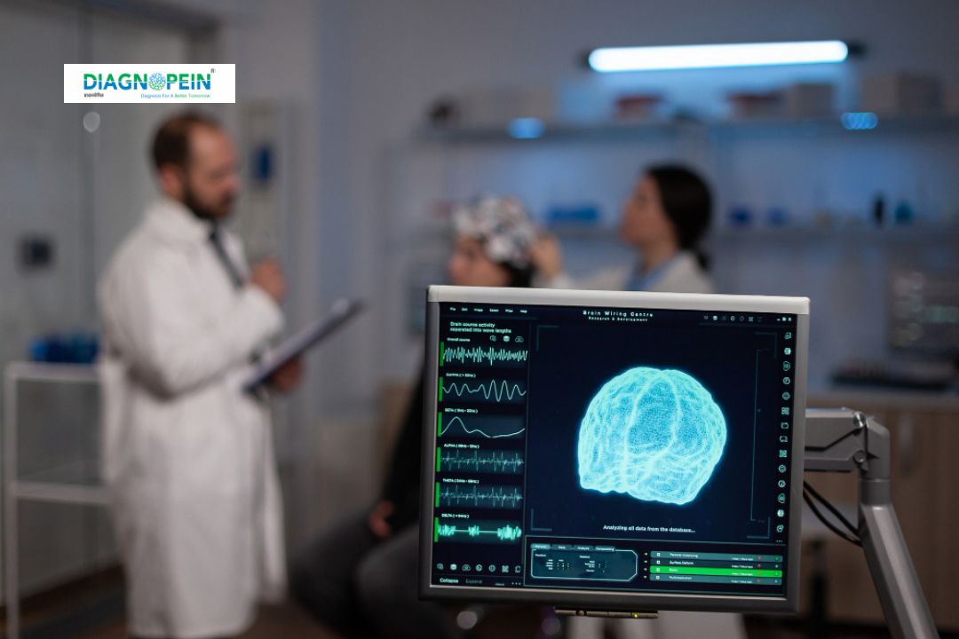
Why MRI Brain Angiography is Needed
MRI Brain Angiography is recommended when symptoms suggest a possible blood flow problem in the brain. It helps identify:
-
Aneurysms or ballooning of brain arteries
-
Arteriovenous malformations (AVMs)
-
Narrowing (stenosis) of vessels causing reduced oxygen supply
-
Stroke, ischemia, or transient ischemic attack (TIA) evaluation
-
Blood vessel inflammation (vasculitis)
-
Abnormal vessel structure near tumors
Unlike CT angiography, MRI Brain Angiography can be done without contrast dye in many cases. This is particularly helpful for patients allergic to iodine-based contrast or those with kidney problems. With rapid scanning techniques, Diagnopein ensures highly accurate reports to guide further treatment or surgical planning.
Benefits of MRI Brain Angiography
MRI Brain Angiography provides several clinical and diagnostic benefits that make it a preferred choice for both patients and neurologists:
-
Non-invasive and radiation-free: It uses magnetic fields instead of X-rays, making it safer for repeated use.
-
High precision: Delivers sharp, 3D images for accurate mapping of blood vessels.
-
Detects early vascular changes: Helps doctors diagnose small vessel abnormalities before they progress.
-
Safe for most patients: Suitable for children and adults; contrast dyes (if used) are much safer than those in CT scans.
-
Comprehensive evaluation: Can also capture soft tissue and brain parenchyma in the same session, giving a full picture of brain health.
Regular MRI Angiography can also monitor patients with a history of high blood pressure, diabetes, or cholesterol issues—factors that increase the risk of cerebrovascular disease.
Testing Procedure and Scan Parameters
Preparation:
Before the scan, patients are asked to remove metal objects and report any implants or pacemakers. Typically, fasting is not required unless contrast material is used. A technologist explains the process to ensure comfort and reduce anxiety.
Procedure:
The test is performed using a 1.5T or 3T MRI machine at Diagnopein. The patient lies on the MRI table, which slides into the scanner. The imaging process lasts about 20–30 minutes. In some cases, a gadolinium-based contrast agent is injected intravenously to enhance vessel visibility.
Key Parameters and Techniques Used:
-
TR/TE: Timing parameters optimized for bright blood imaging (e.g., TOF-MRA).
-
FOV and Slice Thickness: Typically 180–220 mm with 1 mm slices for detailed visualization.
-
Matrix: High-resolution matrix (e.g., 256 x 256) for sharp images.
-
Techniques: Time-of-flight (TOF), Phase-contrast (PC-MRA), or contrast-enhanced MRA depending on patient needs.
After the scan, the radiologist at Diagnopein interprets the images and prepares a comprehensive report. The results help physicians plan medical or surgical interventions precisely and avoid unnecessary procedures.
Why Choose Diagnopein for MRI Brain Angiography
Diagnopein is equipped with cutting-edge MRI systems and a team of expert radiologists specializing in neuroimaging. We ensure accurate, early detection of even minute vascular abnormalities that could impact brain health. Our focus on patient comfort, fast scheduling, and detailed reporting makes us one of the most preferred diagnostic centers for MRI Brain Angiography.
Patients choose Diagnopein because of our:
-
Highly experienced neuro-radiology team
-
Use of 3T MRI scanners for detailed angiographic studies
-
Fast turnaround time with same-day reporting
-
Comfort-oriented patient experience
-
Transparent and affordable pricing
MRI Brain Angiography at Diagnopein ensures peace of mind through reliable, radiation-free diagnostics that allow doctors to make informed therapeutic decisions and patients to take proactive steps toward better neurological health.


