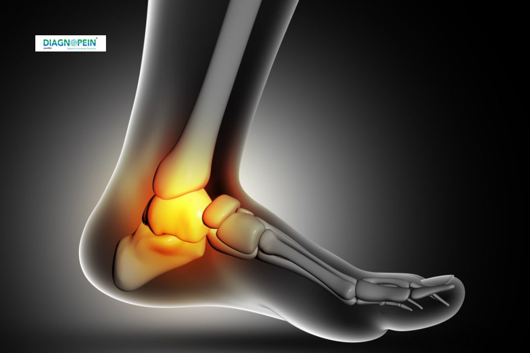
Why MRI Ankle Single Joint – With Contrast is Important
A contrast-enhanced MRI of the ankle provides high-definition imaging, offering deeper insights into complex ankle conditions. It allows detailed visualization of joint inflammation, synovitis, small tears in ligaments or tendons, and early stages of cartilage damage. The contrast material highlights differences in blood flow and tissue composition, making it a preferred diagnostic tool for detecting subtle pathologies.
This test is particularly important for:
-
Detecting early joint infections, abscesses, and inflammation
-
Evaluating ankle ligament injuries and tendon disorders
-
Assessing bone marrow edema, cartilage defects, or small fractures
-
Diagnosing synovial cysts, arthritis, and post-traumatic changes
Timely MRI evaluation supports accurate diagnosis and helps orthopedists, sports medicine experts, and radiologists create a precise treatment plan, avoiding long-term joint damage and mobility loss.
Benefits of MRI Ankle Single Joint – With Contrast
-
High Accuracy: The contrast agent increases image clarity, allowing detection of even minor tissue changes.
-
Comprehensive Joint Assessment: It captures soft tissue, bone, and vascular details in one scan.
-
Non-Invasive Procedure: No radiation exposure; safe for most patients.
-
Early Detection: Identifies hidden causes of ankle pain before they progress to severe conditions.
-
Guided Treatment Planning: Enables accurate decisions for surgery, physiotherapy, or medication.
How the MRI Ankle – With Contrast Test is Done
-
Pre-Procedure Preparation:
The patient is asked to remove all metal objects and change into a hospital gown. Medical history and any known allergies to contrast dye are checked.
-
Contrast Administration:
A gadolinium-based contrast agent is injected through a vein. This dye travels through the bloodstream, highlighting tissues and blood vessels within the ankle.
-
MRI Scanning:
The patient lies still on the MRI table while the machine captures high-resolution images from multiple angles. The scan duration usually lasts 30–45 minutes.
-
Post-Procedure Care:
Most patients can resume normal activities immediately after the scan. The images are sent to a radiologist for interpretation, and results are shared with the referring doctor.
MRI Ankle Single Joint – With Contrast Parameters
-
Magnetic Field Strength: Typically 1.5 or 3 Tesla
-
Sequence Types: T1, T2, STIR, PD-Fat Sat sequences before and after contrast injection
-
Contrast Type: Gadolinium-based agent (as per protocol)
-
Positioning: Patient supine with the ankle in a neutral position
-
Slice Thickness: 2.5–4 mm for detailed evaluation
The scan is safe, but patients with kidney impairment, pregnancy, or known allergic reactions to contrast material should inform their doctor beforehand.
Indications for MRI Ankle – With Contrast
-
Chronic ankle pain or unexplained swelling
-
Sports-related injuries
-
Post-operative evaluation after ligament or tendon repair
-
Suspected bone infections (osteomyelitis)
-
Detection of tumors, cysts, or vascular anomalies