Importance of MRI Ankle Single Joint - Without Contrast
The ankle joint supports your entire body weight and endures significant stress during daily activities. Sports injuries, sprains, chronic pain, or sudden swelling often stem from internal damage that cannot be detected through X-rays or ultrasound alone. MRI Ankle Single Joint - Without Contrast plays a vital role in identifying these soft tissue injuries and joint abnormalities with greater accuracy.
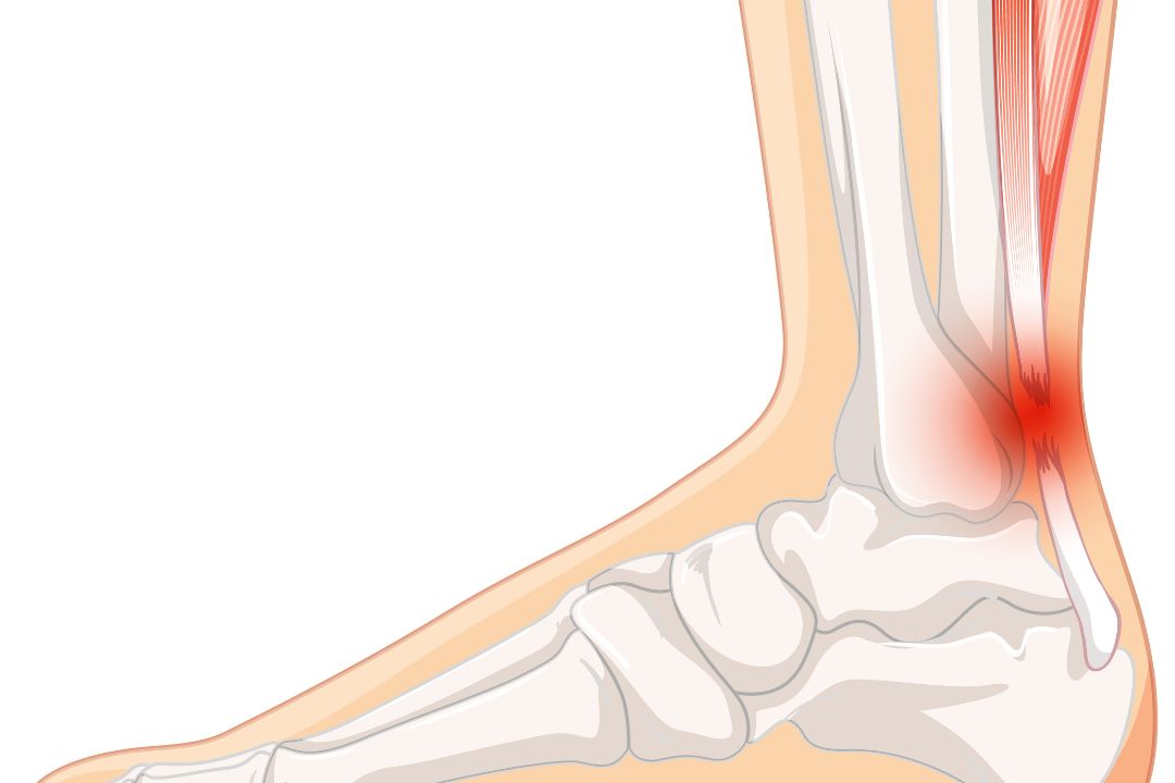
This test helps diagnose:
-
Ligament tears or sprains
-
Tendonitis and tendon ruptures
-
Ankle fractures not visible on X-ray
-
Osteoarthritis and cartilage damage
-
Joint effusion (fluid accumulation)
-
Bone marrow edema and synovial inflammation
By identifying the exact location and extent of injury, this MRI helps your orthopedic or sports medicine specialist decide on the most effective treatment, whether it involves rest, physiotherapy, or surgery.
Benefits of MRI Ankle Single Joint - Without Contrast
MRI is a non-invasive, radiation-free imaging technique that yields detailed 3D views of internal joint structures. Using a non-contrast approach provides additional advantages:
-
No risk of allergic reaction to contrast dye
-
Safe for individuals with kidney disease
-
Short scan duration (20–30 minutes)
-
High accuracy for ligament, tendon, and bone assessment
-
Early detection of inflammatory and degenerative conditions
MRI Ankle Single Joint - Without Contrast offers superior visualization compared to CT scans and X-rays, helping physicians detect early changes before symptoms worsen. The scan’s clarity ensures a faster and more targeted treatment plan, reducing recovery time.
Testing Procedure and Parameters
At Diagnopain, the MRI ankle scan process is comfortable and straightforward. You will be asked to lie down on the scanning table with the target ankle placed in a specialized coil to capture detailed images. No injection or contrast agent is required.
Test Process:
-
The technician positions your foot securely to prevent movement.
-
The MRI machine uses magnetic fields and radio waves to produce images.
-
The scan lasts about 20–30 minutes, during which you must remain still.
-
Images are reviewed by radiologists to detect injuries, tears, or arthritis signs.
Common Parameters Evaluated:
-
Bone marrow signal and alignment
-
Ligaments (anterior talofibular, calcaneofibular, deltoid)
-
Tendons (Achilles, peroneal, posterior tibial)
-
Joint capsule and synovial membrane
-
Cartilage condition and joint space integrity