What is MRI Angiography – With Contrast?
MRI Angiography, commonly known as MRA with contrast, is a diagnostic imaging test that uses strong magnetic fields and radio waves to visualize blood vessels. The contrast agent—typically gadolinium-based—is injected intravenously to enhance visibility of arteries and veins, helping radiologists evaluate circulation and vascular structures clearly.
Unlike conventional angiography, MRA with contrast does not involve ionizing radiation or catheter insertion. This makes it a safer, faster, and more comfortable option for evaluating vascular health.
Why MRI Angiography – With Contrast is Important
MRI Angiography with contrast is crucial for detecting and monitoring conditions related to the circulatory system such as:
-
Brain aneurysms and vascular malformations
-
Carotid artery stenosis or narrowing
-
Peripheral artery disease (PAD)
-
Pulmonary embolism and thoracic vessel abnormalities
-
Renal artery stenosis affecting kidney function
-
Aortic aneurysm and dissection
-
Coronary and cerebral circulation disorders
Early and accurate diagnosis with MRA helps doctors plan effective treatments, surgical interventions, or lifestyle changes to prevent complications such as heart attacks and strokes.
Benefits of MRI Angiography – With Contrast
MRA with contrast offers several key benefits over traditional imaging techniques:
-
Non-invasive procedure: No catheters or surgical tools are required.
-
High-resolution 3D images: Helps visualize complex blood flow patterns with excellent clarity.
-
Radiation-free imaging: Safe for patients who require repeated vascular monitoring.
-
Accurate vessel mapping: Assists surgeons and specialists in treatment planning for angioplasty, stenting, and bypass surgery.
-
Comprehensive evaluation: Detects subtle vascular changes missed by other modalities such as ultrasound or CT.
How the MRI Angiography Test is Done
-
Preparation: There is no special preparation required, though patients may be asked to avoid food or drink for a few hours before the scan. Metallic objects, jewelry, or electronic devices must be removed.
-
Contrast Administration: The radiology technologist administers a gadolinium-based contrast agent through an intravenous (IV) line in the arm.
-
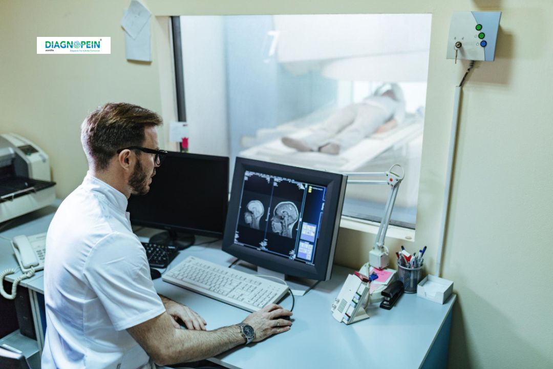
Imaging Process: The patient lies on the MRI table, which slides into the scanner. The magnet creates detailed cross-sectional images as the contrast enhances blood vessel visualization.
-
Duration: The procedure typically lasts 30–60 minutes. The patient must remain still for clear imaging.
-
After the Scan: Patients can usually resume normal activities immediately. Hydration helps flush out the contrast agent naturally through the kidneys.
Parameters Evaluated in MRA with Contrast
MRI Angiography assesses various vascular parameters such as:
-
Blood flow speed and direction
-
Vessel diameter and structural integrity
-
Areas of narrowing, blockage, or aneurysm
-
Arterial wall abnormalities
-
Collateral circulation and perfusion quality
Advanced post-processing tools create 3D reconstructions that allow radiologists to analyze the entire vascular network comprehensively.
Why Choose Diagnopein for MRI Angiography
Diagnopein combines cutting-edge MRI technology, highly skilled radiologists, and a patient-centric approach to deliver accurate vascular diagnostics. Our high-field MRI scanners ensure faster scan times and superior image quality, while experienced technicians ensure comfort and safety during the procedure.
We follow international safety protocols for contrast administration and use only clinically tested agents approved for medical imaging.