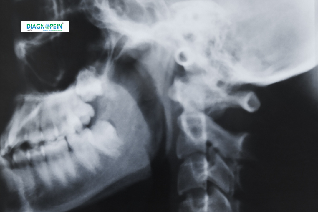
Why MASTOID B/L VIEW is Important
Mastoid air cells can become infected following middle ear infections or sinus problems. When left untreated, these infections may spread deeper into the bone or surrounding regions. A timely Mastoid B/L View helps detect such changes before complications occur.
Key reasons why this test is important:
-
Identifies acute or chronic mastoiditis.
-
Detects sclerosis or loss of air cells in chronic middle ear disease.
-
Helps in evaluating postoperative mastoid cavities.
-
Provides baseline imaging before advanced tests like CT or MRI temporal bone.
-
Guides ENT specialists in planning surgery or long-term treatment.
Early diagnosis through Mastoid B/L X-ray minimizes the risk of complications such as hearing loss, bone abscess, or intracranial spread of infection.
Benefits of Mastoid B/L View
Undergoing this test offers multiple advantages in ear and bone assessment:
-
Quick and simple: The procedure takes only a few minutes to perform.
-
Painless: No discomfort or special preparation is required.
-
Accurate visualization: Provides clear imaging of both mastoid bones simultaneously.
-
Low radiation exposure: Uses minimal X-ray dose compared to CT scans.
-
Affordable and easily available: Widely accessible in diagnostic centers and hospitals.
For patients with persistent ear problems, the Mastoid B/L View is a valuable first-line imaging option that helps identify structural changes efficiently.
How the Test is Performed
The Mastoid B/L View is conducted by a trained radiographer in the X-ray room. The procedure is simple and usually completed in less than 10 minutes.
Step-by-step process:
-
The patient is asked to remove earrings, spectacles, or any metallic items around the head.
-
The patient is positioned with the side of the head aligned against the X-ray plate to capture both mastoid bones.
-
The radiographer adjusts the head tilt and X-ray beam angle to ensure proper alignment.
-
Images are taken from both sides (bilateral view) using a low-dose X-ray beam.
-
The radiologist interprets the images and prepares the report for the referring ENT doctor.
No fasting or contrast medium is needed. Patients can resume normal activities immediately after the test.
Parameters Evaluated in Mastoid B/L View
The test focuses on evaluating key structural and pathological features, including:
-
Size and pattern of mastoid air cells.
-
Evidence of sclerosis or opacification (infection).
-
Bone destruction or erosion.
-
Mucosal thickening and fluid levels.
-
Postoperative changes after mastoidectomy.
-
Symmetry between both mastoid bones.
Interpreting these parameters helps identify disease stages and guides further management.


