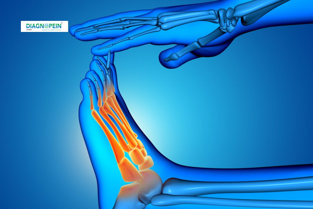
Why LEG AP/LAT. VIEW Is Important
LEG AP and Lateral Views are essential for the assessment of the leg’s structural integrity and pathology. These views help physicians identify bone breaks, dislocations, arthritis, growth disturbances, and other abnormalities in the tibia, fibula, and nearby soft tissues. The AP view offers a silhouette of the leg from front to back, while the lateral view shows the side profile, allowing for comprehensive visual assessment. Accurate diagnosis using LEG AP/LAT. VIEW helps prevent complications, supports timely management, and guides further investigations if needed.
Benefits of LEG AP/LAT. VIEW Imaging
LEG AP/LAT. VIEW X-rays deliver several clinical advantages:
-
Non-invasive, quick, and widely accessible test for leg disorders.
-
High-resolution images that clearly indicate even subtle fractures, bone lesions, and joint malalignment.
-
Immediate results enable fast diagnosis and intervention, benefiting critical cases like trauma or post-accident evaluation.
-
Supports monitoring of bone healing and post-surgical progress.
How LEG AP/LAT. VIEW Testing Is Done
To perform a LEG AP/LAT. VIEW X-ray, the patient is positioned in two ways:
-
For AP view, the patient lies or stands with the leg extended; the X-ray beam passes from front to back of the leg.
-
For lateral view, the patient turns sideways, with the leg positioned laterally; the X-ray beam passes across the leg's side.
Parameters for a high-quality LEG AP/LAT. VIEW include correct positioning, sufficient exposure, and clear visualization of both tibia and fibula, from knee to ankle. Radiation dose is kept minimal, adhering to safety protocols. The images are digitally processed and reviewed by radiologists for diagnosis.


