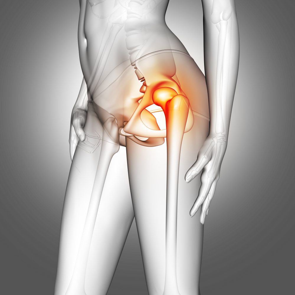
Why HIP AP / LAT. View is Important
The hip joint is one of the strongest weight-bearing joints in the body, making it prone to injuries and wear over time. The HIP AP / LAT. View X-ray is a crucial imaging technique for identifying various conditions, such as:
-
Hip fractures and bone injuries
-
Arthritis and joint degeneration (Osteoarthritis or Rheumatoid Arthritis)
-
Congenital hip dislocation
-
Bone lesions and tumors
-
Pre-surgical and post-operative evaluations
By combining both the AP and Lateral angles, radiologists get an accurate assessment of the bone structures around the pelvis and hip socket (acetabulum). This two-view imaging eliminates the possibility of missing subtle fractures or abnormalities that may not appear clearly in a single projection.
Benefits of HIP AP / LAT. View X-Ray
Performing a HIP AP / LAT. View offers several key benefits:
-
Comprehensive assessment – Provides a complete view of the hip joint from different angles for accurate diagnosis.
-
Non-invasive and quick – The procedure is safe, fast, and does not require anesthesia or contrast.
-
Low radiation exposure – Modern X-ray machines at Diagnopein ensure patient safety with minimal radiation levels.
-
Early detection – Helps detect degenerative joint changes and fractures early for timely treatment.
-
Supports treatment planning – Essential for orthopedic surgeons to plan joint replacement, realignment, or other surgical interventions.
The HIP AP / LAT. View is particularly beneficial for patients who have sustained trauma, hip pain due to aging, or ongoing mobility issues.
How the HIP AP / LAT. View X-Ray is Done
At Diagnopein, the HIP AP / LAT. View procedure is performed by trained radiology professionals following all safety standards.
-
Preparation:
No special preparation is required. The patient may be asked to remove metallic objects near the hip and wear a hospital gown.
-
Positioning:
-
AP View: The patient lies flat on the X-ray table with legs rotated inward to align the hip joint properly.
-
Lateral View: The patient turns slightly on one side to capture the femoral neck and acetabulum clearly.
-
Imaging:
The radiologic technologist positions the X-ray machine above the hip area and captures images from both front and lateral angles.
-
Post-Procedure:
The images are reviewed by a radiologist for diagnosis, and the report is generated usually within a few hours.
Parameters and Technical Details
-
View Type: Anteroposterior (AP) and Lateral (LAT)
-
Purpose: Bone alignment, fracture detection, and degenerative changes
-
Imaging Time: 5–10 minutes
-
Radiation Dose: Low and within diagnostic safety limits
-
Recommended For: Orthopedic trauma, hip pain, arthritis assessment, follow-up after surgery
At Diagnopein, we use digital radiography systems that provide precise, high-resolution HIP AP / LAT. View X-rays, improving diagnostic accuracy and patient comfort.