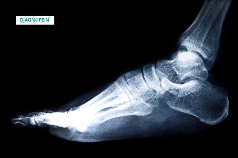
Why is FOOT AP/LAT. VIEW Important?
Foot conditions such as fractures, arthritis, bone spurs, infections, and congenital deformities require precise imaging for accurate diagnosis and effective treatment planning. FOOT AP/LAT. VIEW is critical because it enables doctors to see multiple angles of bones, joints, and soft tissues, helping to:
-
Detect fractures and dislocations with clarity
-
Assess joint spaces in conditions like arthritis
-
Identify bone abnormalities or infections
-
Guide surgical planning and monitor healing progress
Without these views, critical details could be missed, resulting in misdiagnosis or delayed treatment.
Benefits of FOOT AP/LAT. VIEW X-Ray
-
Non-invasive and Quick: The procedure is painless and generally completed within minutes.
-
Cost-effective: Compared to advanced imaging techniques, x-rays are affordable and widely available.
-
High Diagnostic Value: Provides a clear visualization of bone structures, essential for orthopedic and podiatric care.
-
Minimal Radiation Exposure: Uses low-dose radiation making it safe for routine examinations.
These benefits make FOOT AP/LAT. VIEW a default initial diagnostic tool for foot-related complaints.
How is FOOT AP/LAT. VIEW Testing Performed?
The testing involves the following steps:
-
Patient Positioning
-
For the AP view, the patient’s foot is placed flat on the x-ray table or plate with toes pointing upward.
-
For the LAT view, the foot is positioned on its side to capture the profile of the bones.
-
Image Acquisition
-
The radiographer aligns the x-ray beam perpendicular to the foot for the AP view.
-
For the LAT view, the beam is directed from the medial or lateral side depending on clinical requirements.
-
Processing
-
Interpretation
-
The images are analyzed for fractures, dislocations, alignment, bone density, joint integrity, and any abnormal lesions.