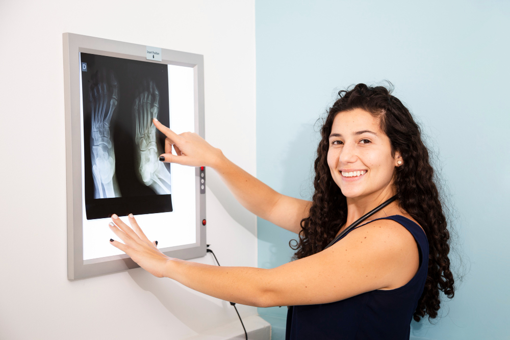
What is a CT Right Wrist Joint Scan?
A CT scan (Computed Tomography) uses X-rays combined with computer technology to create cross-sectional images of your right wrist. Unlike standard X-rays, CT scans show subtle changes in bone and soft tissues, making them vital for cases where physical exams or X-rays don’t reveal enough.
Why Get a CT Scan for the Wrist?
- Unexplained wrist pain not improving with treatment
- Suspected fractures not seen on plain X-rays
- Joint swelling or limited motion after trauma
- Evaluation of old injuries or infections
- Pre-operative planning for wrist surgery
How the Procedure Works
- You’ll rest your right arm on a scanning table.
- The technologist positions your wrist; sometimes, a mild contrast dye may be injected for better joint visualization.
- The scanner rotates around your wrist, capturing multiple images in just a few minutes.
- No pain, minimal discomfort, and results are quickly available.
Key Benefits
- Rapid, detailed visualization—perfect for urgent injury cases
- Pinpoints even tiny fractures or bone fragments
- Guides orthopedic or surgical planning with clarity
- No hospital stay; done in outpatient settings
What to Expect After the Scan
You can return to daily activities within minutes. Your doctor will review images and explain the findings, whether it’s a simple sprain or something more complicated. Advanced imaging makes treatment easier and outcomes better.
Tips for Best Results
- Wear comfortable clothing and remove metal accessories.
- Inform the technologist about allergies, pregnancy, or previous procedures.
- Arrive early to relax and understand the process.
Why Choose a CT Scan?
CT right wrist joint scans are the gold standard for precision. Fast, clear, and exceptionally accurate—so you get answers and relief faster. Talk with your healthcare provider about whether CT is right for you.


