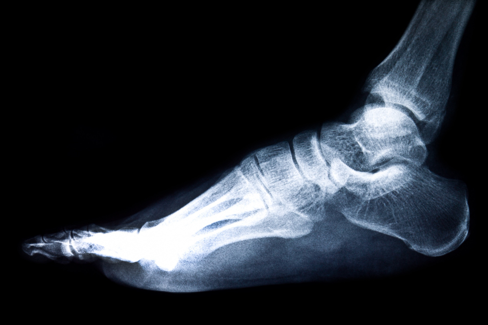
Why the Test Is Done
Doctors recommend a CT Right Foot for:
- Unexplained or persistent right foot pain
- Evaluation of suspected hairline or complex fractures
- Detection of arthritis, bone infections, or tumors
- Planning before orthopedic surgeries
- Monitoring healing after injury or surgery
The scan offers high diagnostic accuracy, guiding faster and more effective treatment.
How the Procedure Works
- You’ll be asked to remove footwear and any metal items.
- The technologist will position your right foot on the CT table.
- The scanner takes a series of X-ray images from multiple angles.
- These images are combined to create a 3D view of your foot structures.
The process is quick—typically completed within 10–15 minutes—and entirely painless. In some cases, contrast dye may be used to highlight soft tissues or blood vessels.
Benefits of CT Right Foot Imaging
- Fast and accurate diagnosis of bone and soft-tissue injuries
- 3D visualization helping doctors plan surgeries precisely
- Early detection of complex fractures and degenerative changes
- Non-invasive and comfortable for patients
- Improved recovery outcomes through timely treatment decisions
Preparing for the Scan
No special preparation is needed in most cases. If contrast dye is required, you might be asked to avoid eating for a few hours prior. Always inform your doctor about allergies, pregnancy, or existing medical conditions.
Why Choose a CT Right Foot Scan
Choosing a CT scan ensures you get the most detailed view of your right foot structures, supporting accurate diagnosis and targeted treatment. Whether it’s a sports injury, chronic pain, or post-operative review, this test helps clinicians detect and manage conditions effectively.