


Diagnopein CT LT KNEE JOINT Centre in Karad
Diagnopein CT LT KNEE JOINT Centre in Karad
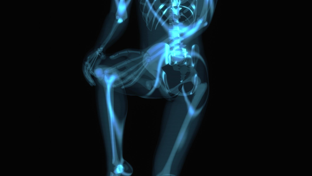
A CT LT Knee Joint scan is a specialized imaging procedure that provides detailed cross-sectional images of the left knee, focusing on the bones, joint structures, and surrounding tissues. It is primarily used to detect fractures, bone lesions, degenerative changes such as arthritis, and other abnormalities in the knee that may not be clearly visible on standard X-rays. This scan is particularly useful for preoperative planning, assessing postoperative healing, and evaluating cases where MRI is contraindicated, especially when metal implants are present.
The procedure involves the patient lying supine while a CT scanner captures thin-slice images of the distal femur, proximal tibia, patella, and fibular head.


