Why CT Temporal Bone Is Important
A CT Temporal Bone scan plays a critical role in diagnosing and managing ear and skull-base disorders. Because the temporal bone contains delicate and tightly packed structures, even small abnormalities can cause significant symptoms such as hearing loss, vertigo, tinnitus, or facial weakness.
Doctors often recommend this scan to detect an abnormal CT scan temporal bone when patients present with chronic ear discharge, repeated ear infections, dizziness, or unexplained hearing impairment. It is also crucial in pre-operative planning and post-surgical assessment.
Conditions evaluated using CT of mastoid bone and CT petrous temporal bone include:
-
Chronic otitis media and cholesteatoma
-
Mastoiditis and bone erosion
-
Congenital ear malformations
-
Temporal bone fractures
-
Inner ear abnormalities
-
Facial nerve canal involvement
At Diagnopein Ahmednagar, our radiologists specialize in CT temporal bone radiology, ensuring accurate interpretation for timely treatment.
Benefits of CT Temporal Bone Scan
Choosing a CT Temporal Bone scan offers several important advantages:
-
High-Resolution Imaging
The scan provides extremely detailed images of the middle ear ossicles, mastoid air cells, cochlea, semicircular canals, and petrous bone.
-
Early and Accurate Diagnosis
Conditions such as cholesteatoma, mastoid infection, and bone destruction can be detected early through CT mastoid scan and CT mastoid bone imaging.
-
Surgical Planning Support
ENT surgeons rely on CT temporal bone images to plan complex ear surgeries safely and precisely.
-
Contrast and Non-Contrast Options
Depending on clinical needs, CT temporal bone with contrast or CECT temporal bone helps evaluate soft tissue involvement, tumors, and vascular abnormalities.
-
Quick and Non-Invasive
The scan is painless, takes only a few minutes, and does not require hospital admission.
At Diagnopein Diagnostic Centre, Ahmednagar, we ensure patient comfort, safety, and accurate reporting.
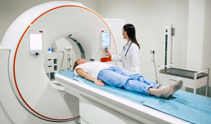
How CT Temporal Bone Testing Is Done
The CT temporal scan procedure is simple and well-tolerated:
-
The patient lies comfortably on the CT table.
-
The head is positioned carefully to focus on the temporal bone region.
-
The CT scanner rotates around the head to capture thin-slice images.
-
For CECT temporal bone, a contrast dye may be injected intravenously if advised by the doctor.
-
The entire scan usually takes 10–15 minutes.
Patients are advised to remain still during the scan to obtain clear CT temporal bone images. No special preparation is required unless contrast is used.
CT Temporal Bone Scan Parameters
At Diagnopein Ahmednagar, standardized protocols are followed to ensure optimal image quality:
-
Slice thickness: 0.5–1 mm
-
Scan plane: Axial and coronal reconstructions
-
Field of view: Focused temporal bone region
-
Window settings: Bone and soft tissue windows
-
Contrast usage: Only for CT temporal bone with contrast when clinically indicated
These parameters help in detailed evaluation of CT anatomy of temporal bone, mastoid air cells, and petrous structures.
Why Choose Diagnopein Diagnostic Centre, Ahmednagar
-
Advanced multi-slice CT technology
-
Expert radiologists in CT temporal bone radiology
-
Accurate and timely reports
-
Affordable CT temporal bone cost
-
Patient-friendly environment
-
Trusted diagnostic center in Ahmednagar