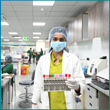
The cell block test is a critical laboratory procedure used primarily in the field of cytopathology to analyze and diagnose various diseases, particularly cancers


The cell block test involves the preparation of a solid tissue sample from a fluid specimen, such as a fine-needle aspiration (FNA) or other body fluids like pleural effusions, ascites, or bronchial washings. The test is designed to create a more structured and representative sample for microscopic examination, enabling pathologists to evaluate the cellular characteristics and morphology of the specimen.
1] Diagnostic Tool
The primary purpose of the cell block test is to aid in the diagnosis of various medical conditions, particularly:
A] Cancer: The cell block test is crucial in identifying malignancies, as it allows for the evaluation of cellular features such as size, shape, and arrangement.
B] Infections: It can help detect infectious agents by examining the cellular response and identifying abnormal cells.
C] Inflammatory Conditions: The test is useful in assessing inflammatory processes, helping to differentiate between benign and malignant conditions.
2] Research Applications - In addition to clinical diagnostics, cell blocks are used in research settings to study disease mechanisms, cellular behavior, and responses to treatment, making them an important tool in both clinical and academic laboratories.
1] Sample Collection - The first step in the cell block procedure involves collecting the sample. Common methods include:
A] Fine-Needle Aspiration (FNA): A thin needle is used to extract cells from a suspicious mass or lesion.
B] Body Fluid Collection: Fluids such as pleural effusions or peritoneal fluid are collected for analysis.
2] Processing the Sample - Once the sample is obtained, the following steps are typically followed:
A] Centrifugation: The fluid specimen is centrifuged to separate cells from the supernatant, concentrating the cellular components.
B] Embedding: The sediment containing the cells is then embedded in a fixative, usually paraffin wax or agar, to create a solid block that preserves the cellular architecture.
C] Sectioning: Thin sections of the block are cut using a microtome, allowing pathologists to examine the sample under a microscope.
D] Staining: The sections are stained using various dyes, such as Hematoxylin and Eosin (H&E), to enhance visibility and contrast of cellular components.
3] Microscopic Examination - The final step involves examining the stained sections under a microscope. Pathologists analyze the cellular morphology, looking for abnormal features that may indicate disease.
You should choose best diagnostic centre in Nagpur for your CELL BLOCK test because we are committed to providing high-quality diagnostic care in a clean and hygienic environment. Our center is equipped with advanced technology to ensure accurate and reliable test results, which are crucial for assessing this test. Diagnopein’s experienced staff is dedicated to handling tests with precision and care, offering you both expertise and comfort. We also offer affordable pricing, making essential health diagnostics accessible without compromising quality. For timely, professional, and affordable cardiac testing, Diagnopein is your trusted partner for your healthcare.
Common samples include those collected from fine-needle aspirations, pleural effusions, ascites, bronchial washings, and other body fluids.
Fasting is generally not required, but specific instructions may vary based on the clinical context and the physician's recommendations.
This test is performed to diagnose diseases such as cancer, infections, and inflammatory conditions. It provides more structured samples for pathologists to analyze compared to traditional cytology.
Yes, limitations include potential sample quality issues, a more time-consuming process compared to direct cytology, and the need for specialized training and equipment.